

Why Do My Stomach Hurt Every Time I Eat? Causes, Symptoms, and What to Do
September 3, 2026
Where Can I Find Dental Offices Hiring Near Me and Get Hired?
September 3, 2026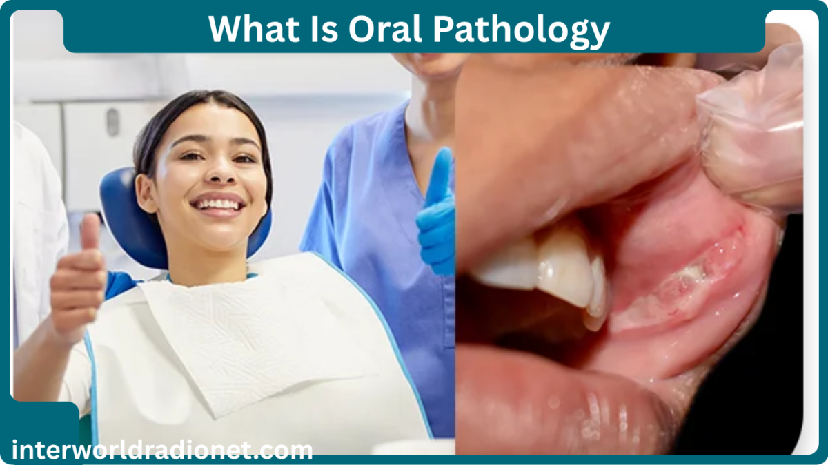
Oral pathology is the branch of dentistry and pathology that focuses on diseases and abnormal conditions affecting the mouth, jaws, salivary glands, and related tissues. It helps explain why a person may develop a mouth sore, white patch, red area, swelling, lump, unusual pigmentation, jaw lesion, or other change inside or around the mouth.
The mouth can show signs of many different problems. Some are temporary and harmless, while others need treatment. In some cases, changes in the mouth can also provide clues about a health problem elsewhere in the body.
Oral pathology is therefore much more than looking for oral cancer. It covers a very broad range of conditions, including infections, inflammatory diseases, immune-related disorders, cysts, benign growths, potentially precancerous lesions, cancer, salivary gland disorders, and diseases involving the jawbones.
A major challenge is that different oral conditions can look surprisingly similar. A white patch may result from irritation, infection, a benign change, or a potentially serious lesion. A lump may be harmless, but a persistent lump should still be evaluated. A mouth ulcer may be a common canker sore, but an ulcer that does not heal needs professional attention.
This is why oral pathology relies on careful clinical examination and, when necessary, laboratory testing and biopsy rather than appearance alone. A biopsy allows a pathologist to examine tissue under a microscope and determine what is happening at the cellular level.
This comprehensive guide explains oral pathology in simple language, including what an oral pathologist does, common oral diseases, causes of oral lesions, diagnostic methods, oral cancer, treatment options, warning signs, and questions patients commonly ask.
What Is Oral Pathology?
Oral pathology is the medical and dental study of diseases affecting the oral and maxillofacial region.
The word “oral” refers to the mouth, while “pathology” refers to the study of disease and abnormal changes in tissues.
Depending on the condition, oral pathology may involve:
- Lips
- Tongue
- Gums
- Inner cheeks
- Roof of the mouth
- Floor of the mouth
- Tonsillar region
- Salivary glands
- Jawbones
- Oral mucous membranes
- Soft tissues around the mouth
The oral cavity includes several specific anatomical areas, including the lips, front portion of the tongue, gums, inner cheeks, floor of the mouth, hard palate, and related structures.
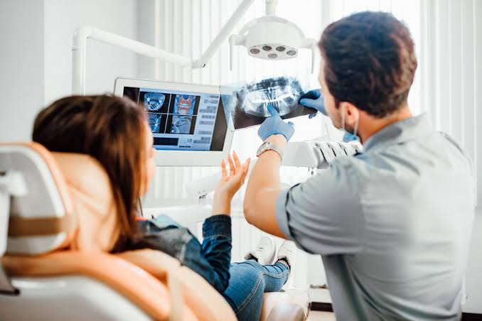
An oral pathologist is a specialist who studies diseases affecting these areas. They may examine tissue removed during a biopsy and prepare a pathology report that helps the dentist, oral surgeon, physician, or other healthcare professional determine the diagnosis.
Also read:What Is an Odontologist? A Complete Guide to Forensic Dentistry, Training, Jobs, and What They Do
What Does an Oral Pathologist Do?
An oral pathologist helps identify diseases by studying abnormal tissues, cells, and clinical findings.
A patient may never meet an oral pathologist directly. In many cases, a dentist or oral surgeon notices an abnormal area, removes a small tissue sample, and sends it to a pathology laboratory.
The oral pathologist then examines the specimen.
The evaluation may involve:
- Examining the tissue with the naked eye
- Processing the tissue
- Preparing microscopic slides
- Studying cells under a microscope
- Looking for inflammation
- Identifying infection
- Assessing abnormal cell growth
- Looking for precancerous changes
- Determining whether cancer is present
- Providing a final pathology diagnosis
The pathology report can then help the treating clinician decide what should happen next.
This is one reason oral pathology is so important. A clinical examination may suggest several possible diagnoses, but microscopic examination can provide much more specific information.
Why Is Oral Pathology Important?
Your mouth is constantly exposed to food, bacteria, temperature changes, chemicals, physical pressure, and other environmental factors.
At the same time, oral tissues can be affected by medications, infections, immune conditions, nutritional problems, systemic diseases, and genetic disorders.
Because so many factors can affect the mouth, oral changes can have many explanations.
Oral pathology helps separate these possibilities.
For example, suppose someone develops a persistent white patch inside the cheek.
The patch could be caused by:
- Repeated friction
- Cheek biting
- Tobacco exposure
- A fungal infection
- An inflammatory condition
- A potentially malignant lesion
- Another oral disorder
Simply looking at the patch may not always be enough.
A trained dental professional can evaluate its location, texture, duration, appearance, and other characteristics and determine whether additional testing is necessary.
What Conditions Does Oral Pathology Cover?
Oral pathology includes a very large group of diseases.
These can generally be organized into several categories:
- Oral infections
- Inflammatory conditions
- Ulcers
- White lesions
- Red lesions
- Pigmented lesions
- Cysts
- Benign tumors
- Salivary gland disorders
- Jawbone diseases
- Immune-related disorders
- Developmental abnormalities
- Potentially malignant disorders
- Oral cancers
Many oral lesions are benign. However, some require monitoring or treatment because they can represent a higher-risk change.
MSD Manual notes that growths in and around the mouth may be benign, precancerous, or malignant, with most mouth growths being noncancerous.
Common Oral Pathology Conditions
There is no single “typical” oral pathology condition.
Different diseases produce very different symptoms.
Some cause pain. Others cause no symptoms at all.
Some disappear within days. Others remain for weeks or months.
Understanding the major categories can help you recognize why professional examination matters.
Mouth Ulcers and Canker Sores
Mouth ulcers are among the most common oral problems.
A canker sore, medically called recurrent aphthous stomatitis, is usually a painful ulcer that develops inside the mouth.
It may occur on areas such as:
- Inner lips
- Inner cheeks
- Tongue
- Soft oral tissues
Most ordinary canker sores eventually heal.
However, not every mouth ulcer is a canker sore.
Ulcers can also result from:
- Physical injury
- Infections
- Certain medications
- Nutritional deficiencies
- Immune disorders
- Systemic diseases
- Chemical irritation
- Other oral conditions
Mouth sores can have infectious, inflammatory, traumatic, allergic, medication-related, or systemic causes.
This is why a persistent ulcer should not automatically be dismissed as a canker sore.
Oral Thrush
Oral thrush is a fungal infection involving the mouth.
It is commonly associated with an overgrowth of Candida species.
Thrush may produce whitish patches that can resemble soft or creamy material on the oral lining.
It can occur more often in certain situations, including:
- Recent antibiotic use
- Corticosteroid use
- Reduced immune function
- Certain medical conditions
The appearance can sometimes be distinctive, but a professional examination is still useful because other white oral lesions can have different causes.
MSD Manual notes that Candida normally exists in the mouth but can overgrow in certain circumstances, producing oral candidiasis.
White Oral Lesions
White patches are a major area of interest in oral pathology.
A white area may develop because the tissue becomes thicker, irritated, infected, or otherwise altered.
Possible causes include:
- Chronic irritation
- Cheek biting
- Tobacco exposure
- Fungal infection
- Lichen planus
- Leukoplakia
- Other mucosal conditions
Some white areas can be wiped away, while others cannot.
That difference can provide useful information, but it does not always establish a diagnosis.
Leukoplakia is a clinical term used for a white patch or plaque that cannot be otherwise characterized as another disease. Some leukoplakic lesions can have potentially malignant changes, which is why persistent unexplained white patches should be professionally evaluated.
Red Oral Lesions
Red areas in the mouth also deserve attention.
An unexplained red patch may have many possible causes.
One important condition is erythroplakia, which describes a red, flat, or worn-looking area of oral mucosa that cannot be explained by another diagnosis.
Red lesions can sometimes have a greater concern for abnormal cellular changes than ordinary white lesions.
MSD Manual describes erythroplakia as a potentially concerning oral lesion and notes that it can be associated with oral cancer.
This does not mean every red area is cancer.
It means that persistent unexplained red lesions should not be ignored.
Mixed Red and White Oral Lesions
Some oral lesions contain both red and white areas.
These are sometimes called mixed red-white lesions.
A mixed appearance can occur in different conditions, but persistent mixed lesions deserve careful evaluation because certain potentially malignant disorders may appear this way.
The exact diagnosis may require a biopsy.
This is another example of why color alone cannot provide a final diagnosis.
Oral Lichen Planus
Oral lichen planus is an inflammatory condition that can affect the lining of the mouth.
It may produce white, lace-like patterns, particularly on the inside of the cheeks.
Some people have no major symptoms.
Others may experience:
- Burning
- Soreness
- Sensitivity
- Pain with spicy foods
- Red or eroded areas
The condition can vary greatly from person to person.
Because some forms can resemble other oral diseases, professional evaluation is important.
MSD Manual describes oral lichen planus as a condition that may produce a lacy pattern known as Wickham striae and may sometimes become erosive.
Oral Herpes and Other Viral Infections
Viruses can cause oral lesions.
Herpes simplex virus is a well-known example.
Cold sores often appear around the lips, but herpes infections can also affect oral tissues.
Other viruses may produce mouth sores as part of a wider illness.
For example, some viral infections can cause multiple sores together with fever, rash, or other symptoms.
The number, location, appearance, and timing of lesions can help a clinician identify the likely cause.
Traumatic Oral Lesions
Not every mouth lesion represents disease.
Sometimes the tissue is simply injured.
Common causes include:
- Accidentally biting the cheek
- Sharp teeth
- Broken fillings
- Dental appliances
- Poorly fitting dentures
- Aggressive brushing
- Very hot food
- Chemical irritation
For example, very hot food can burn the roof of the mouth.
A damaged area may then become painful and develop an ulcer.
If the source of irritation is removed, the tissue may heal.
But if the lesion remains after the suspected cause has been corrected, it should be examined.
MSD Manual notes that physical injury and irritation from teeth, dentures, foods, chemicals, and other sources can produce oral blisters or ulcers.
Salivary Gland Disorders
Saliva plays an important role in oral health.
It helps lubricate the mouth, assists swallowing, supports digestion, and provides protection to oral tissues.
Problems involving the salivary glands can therefore produce several symptoms.
These may include:
- Dry mouth
- Swelling
- Pain near a gland
- Difficulty eating
- Recurrent infections
- Changes in saliva flow
Salivary gland disorders may involve infections, blocked ducts, cysts, stones, inflammation, or tumors.
The location of swelling can help a clinician determine which gland may be involved.
Dry Mouth and Oral Pathology
Dry mouth, medically known as xerostomia, can affect oral health in several ways.
Saliva normally protects the mouth.
When saliva production decreases, the oral tissues may become more vulnerable to irritation, infection, tooth decay, and mouth sores.
Dry mouth may be associated with:
- Certain medications
- Dehydration
- Some medical conditions
- Radiation treatment
- Salivary gland problems
- Other causes
Because reduced saliva can make oral tissues more vulnerable, persistent dry mouth should not be ignored.
MSD Manual specifically notes that reduced saliva production can increase the likelihood of mouth sores and inflammation.
Oral Cysts
An oral cyst is a closed sac or cavity that may contain fluid, tissue, or other material.
Cysts can occur in soft tissues or within the jawbone.
Some are related to teeth.
Others develop from developmental or other processes.
Depending on their size and location, cysts may cause:
- Swelling
- Pressure
- Tooth displacement
- Jaw expansion
- Pain
- No symptoms at all
Some cysts are discovered accidentally during dental X-rays.
This is one reason routine dental imaging can sometimes reveal conditions that a patient cannot feel.
Benign Oral Growths
Many oral growths are noncancerous.
They may develop from:
- Connective tissue
- Bone
- Muscle
- Nerve tissue
- Blood vessels
- Salivary tissues
- Other structures
Examples include certain fibromas, lipomas, vascular lesions, and other benign growths.
A benign growth can still cause problems if it becomes large, repeatedly injured, affects eating, or interferes with speech.
Most importantly, a new or persistent lump should be evaluated rather than self-diagnosed.
Jawbone Lesions
Oral pathology also includes diseases affecting the bones of the jaw.
Some jaw lesions are cystic.
Others are tumors, inflammatory conditions, developmental abnormalities, or other bone disorders.
Possible symptoms include:
- Jaw swelling
- Facial asymmetry
- Tooth movement
- Numbness
- Pain
- Changes in the bite
- Delayed tooth eruption
- A lesion seen on an X-ray
Some jaw lesions cause no symptoms and are discovered during routine dental imaging.
Depending on the findings, a dentist may refer the patient to an oral and maxillofacial surgeon or other specialist.
Oral Cancer and Oral Pathology
Oral cancer is one of the most important reasons persistent oral lesions require evaluation.
Most oral cavity cancers are squamous cell carcinomas, which develop from squamous cells lining areas of the oral cavity.
Oral cavity cancer can occur in locations including:
- Lips
- Front portion of the tongue
- Gums
- Inner cheeks
- Floor of the mouth
- Hard palate
The appearance of oral cancer can vary.
It may resemble:
- A persistent ulcer
- A white patch
- A red patch
- A mixed red-white lesion
- A lump
- An area of thickening
- A nonhealing wound
This is why visual appearance alone cannot reliably distinguish cancer from a harmless condition.
What Are the Risk Factors for Oral Cancer?
Several factors can increase oral cancer risk.
Important risk factors include tobacco use and alcohol consumption.
Using both tobacco and alcohol can create an even greater risk than either exposure alone.
Certain oral HPV infections are also associated with cancers in the oropharyngeal region, particularly cancers involving areas such as the tonsils and base of the tongue.
Risk does not mean that a person will definitely develop cancer.
Likewise, a person without obvious risk factors can still develop an oral malignancy.
That is why persistent changes should be evaluated based on the lesion itself rather than risk factors alone.
What Oral Cancer Warning Signs Should You Know?
Potential warning signs include:
- A mouth sore that does not heal
- An unexplained lump
- Persistent white patches
- Persistent red patches
- Red and white mixed areas
- Unexplained bleeding
- Persistent pain
- Difficulty swallowing
- Difficulty chewing
- Persistent numbness
- A change in how dentures fit
- Unexplained loose teeth
- A persistent sore throat
- A change in voice
- A lump in the neck
These symptoms do not automatically mean cancer.
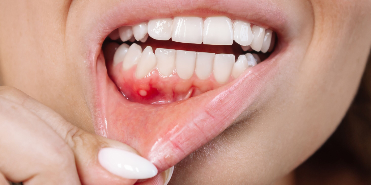
Many have much more common explanations.
However, persistence is important.
An oral lesion that repeatedly returns, does not heal, grows, bleeds, or changes should be examined.
Also read:What Can Dentists Do? A Complete Guide to Dental Treatments and Services
Why Does a Biopsy Matter in Oral Pathology?
A biopsy is one of the most important diagnostic tools in oral pathology.
During a biopsy, a healthcare professional removes a sample of abnormal tissue.
The tissue is sent to a laboratory.
A pathologist then examines it under a microscope.
The report may determine whether the tissue shows:
- Normal changes
- Inflammation
- Infection
- Benign growth
- Dysplasia
- Precancerous changes
- Cancer
- Another specific disease
The National Cancer Institute explains that a biopsy involves removing tissue or cells so a pathologist can examine them under a microscope and make or confirm a diagnosis.
What Happens During an Oral Biopsy?
The exact procedure depends on the location and size of the lesion.
A small lesion may sometimes be removed entirely.
This is called an excisional biopsy.
If a lesion is larger, only part of it may be removed.
This is called an incisional biopsy.
Local anesthesia is commonly used so the area is numb during the procedure.
Afterward, the tissue is sent to a pathology laboratory.
The patient may receive instructions about eating, brushing, pain control, and wound care.
The pathology report may take several days or longer depending on the complexity of the specimen and whether additional testing is required.
Are Oral Lesions Always Biopsied?
No.
Not every oral lesion needs a biopsy.
A dental professional may diagnose some common conditions based on history and clinical appearance.
For example, a lesion with a clear cause that heals normally may not require tissue sampling.
A biopsy becomes more important when the diagnosis is uncertain or when the lesion is persistent, suspicious, changing, or potentially precancerous.
The decision depends on factors such as:
- Location
- Size
- Duration
- Color
- Texture
- Symptoms
- Patient history
- Risk factors
- Changes over time
What Is a Pathology Report?
A pathology report is a medical document describing what the pathologist found in the tissue.
It may include:
- The type of tissue examined
- The diagnosis
- Microscopic findings
- Degree of abnormality
- Whether cancer is present
- Whether margins are involved when appropriate
- Additional testing
- Other important microscopic details
Pathology reports can sometimes contain technical language.
Patients should not feel embarrassed about asking their dentist or doctor to explain unfamiliar terms.
Understanding the diagnosis is an important part of making informed healthcare decisions.
What Is Dysplasia in Oral Pathology?
Dysplasia refers to abnormal changes in cells or tissue.
Oral epithelial dysplasia can occur in certain potentially malignant lesions.
Dysplasia does not automatically mean cancer.
Instead, it describes abnormal cellular changes that can be associated with an increased risk of developing cancer.
The significance depends on factors such as the severity and characteristics of the lesion.
This is why pathology terminology can be confusing.
“Abnormal” does not always mean “cancer.”
How Is Oral Pathology Diagnosed?
Diagnosis usually begins with a detailed history and oral examination.
The clinician may examine:
- Lips
- Tongue
- Gums
- Cheeks
- Palate
- Floor of the mouth
- Teeth
- Jaw
- Neck
They may also feel areas of swelling or enlarged lymph nodes.
Depending on the problem, additional tests may be necessary.
These can include:
- Dental X-rays
- Panoramic imaging
- CT scans
- MRI
- Blood tests
- Microbiological testing
- Saliva-related testing
- Biopsy
- Microscopic examination
The best test depends on the suspected condition.
Can Dental X-Rays Detect Oral Pathology?
Dental imaging can reveal many problems that cannot be seen during an ordinary visual examination.
X-rays can help identify:
- Jaw cysts
- Bone lesions
- Tooth-related infections
- Abnormal bone changes
- Impacted teeth
- Other hidden abnormalities
More advanced imaging, such as CT or MRI, may be used when additional detail is needed.
However, imaging does not replace tissue diagnosis when a lesion requires biopsy.
What Is the Difference Between an Oral Pathologist and a General Dentist?
A general dentist provides broad dental care, including examinations, prevention, fillings, crowns, gum care, and many other services.
An oral pathologist specializes in diagnosing diseases of the mouth and related structures, particularly through examination of tissues and microscopic pathology.
A general dentist may be the first person to notice an abnormal lesion.
The dentist may then refer the patient to an oral pathologist, oral surgeon, ENT specialist, dermatologist, or another professional depending on the suspected problem.
This team-based approach is often important because oral diseases can cross the boundaries of dentistry and medicine.
Can Oral Pathology Reveal a Systemic Disease?
Yes.
The mouth can sometimes show signs associated with health problems elsewhere in the body.
Examples of systemic conditions that may produce oral findings include:
- Nutritional deficiencies
- Blood disorders
- Immune disorders
- Gastrointestinal diseases
- Certain infections
- Endocrine conditions
- Genetic disorders
For example, some nutritional deficiencies can be associated with changes in the tongue or mouth sores. Certain systemic diseases can also produce characteristic oral findings.
This does not mean an oral symptom automatically identifies a systemic disease.
Instead, oral findings can sometimes provide an important clue that encourages further evaluation.
How Does Tobacco Affect Oral Health?
Tobacco can damage oral tissues in several ways.
It can contribute to irritation, changes in the oral lining, gum disease, tooth problems, and increased oral cancer risk.
Tobacco exposure may also contribute to certain oral lesions.
The risk is relevant to both smoked and smokeless tobacco products.
Stopping tobacco use is one of the most important steps a person can take to protect oral health.
Can Alcohol Affect Oral Pathology?
Heavy alcohol use is an established risk factor for several head and neck cancers.
The risk becomes particularly concerning when alcohol use occurs together with tobacco exposure.
Reducing alcohol intake can therefore be an important part of lowering preventable oral cancer risk.
If you are concerned about your personal risk, a dentist or healthcare professional can help you understand which factors are most relevant to you.
Can HPV Affect the Mouth?
Certain types of human papillomavirus can infect oral and throat tissues.
HPV is particularly important in relation to oropharyngeal cancers.
HPV-associated cancers often involve areas such as the tonsils and base of the tongue rather than the front part of the oral cavity.
The presence of HPV does not mean that a person has cancer.
Most HPV infections do not result in cancer.
The important point is that HPV is one of several factors involved in head and neck disease.
How Can You Protect Your Oral Health?
Good oral health begins with consistent everyday habits.
Important steps include:
- Brush your teeth regularly.
- Clean between your teeth.
- Visit your dentist as recommended.
- Avoid tobacco.
- Limit excessive alcohol.
- Eat a balanced diet.
- Address persistent dry mouth.
- Protect your mouth during contact sports.
- Do not ignore persistent oral lesions.
- Follow up when your dentist recommends additional testing.
Prevention cannot eliminate every oral disease.
However, it can reduce many common dental problems and improve the chance of identifying abnormalities early.
Why Regular Dental Examinations Matter
Dental appointments are not only about cavities.
A dental professional can examine the soft tissues of your mouth and look for unusual changes.
The examination may include checking the:
- Tongue
- Cheeks
- Lips
- Gums
- Palate
- Floor of the mouth
- Jaw
- Neck
Oral examinations can identify lesions that patients may not notice themselves.
The National Cancer Institute notes that dentists and physicians may examine the oral cavity for lesions such as leukoplakia and erythroplakia.
What Should You Do If You Find a New Mouth Lesion?
Do not panic.
Many mouth lesions are harmless.
Instead, pay attention to what happens next.
Consider:
- When did it begin?
- Is it painful?
- Is it getting bigger?
- Is it changing color?
- Does it bleed?
- Did you recently injure the area?
- Did you change toothpaste or mouthwash?
- Did you start a new medication?
- Does it heal normally?
If the lesion persists or changes, arrange a dental or medical evaluation.
Taking a photograph can sometimes help you compare how the lesion changes over time, but a photograph should not replace an examination.
Why Self-Diagnosing Oral Lesions Can Be Risky
Searching for pictures online can be useful for general education, but it has limits.
Many oral conditions look alike.
A white patch may represent a harmless condition in one person and require further testing in another.
A mouth ulcer may be a common canker sore or may have another cause.
A lump may be benign, but it still needs evaluation if it persists.
Even professionals sometimes need biopsy results to make a definite diagnosis.
This is why oral pathology combines clinical examination with laboratory evidence when appropriate.
What Treatments Are Used in Oral Pathology?
Treatment depends entirely on the diagnosis.
Possible treatments include:
- Removing an irritant
- Antifungal medication
- Antiviral treatment when appropriate
- Anti-inflammatory treatment
- Dental restoration
- Treatment of gum disease
- Cyst removal
- Surgical removal of a benign growth
- Treatment of an underlying systemic condition
- Monitoring of a low-risk lesion
- Surgical treatment of cancer
- Radiation therapy
- Chemotherapy
- Immunotherapy
- Targeted therapy
Not every lesion requires treatment.
Also read:What Is a DMD? Meaning, Degree, Education, Licensing, and Dental Career Guide
Some only require observation.
Others need immediate intervention.
The pathology diagnosis helps guide that decision.
What Is the Most Important Lesson About Oral Pathology?
The biggest lesson is that persistence matters.
A single sore after accidentally biting your cheek is usually very different from an unexplained lesion that remains for weeks.
A temporary irritation is different from a growing lump.
A normal variation is different from a changing red or white patch.
The longer an unexplained abnormality remains, the more important professional evaluation becomes.
This does not mean that a persistent lesion is necessarily cancer.
It means that the cause should be established rather than guessed.
Frequently Asked Questions About Oral Pathology
Is oral pathology only about oral cancer?
No. Oral pathology covers many conditions besides cancer, including infections, ulcers, inflammatory diseases, cysts, benign growths, salivary gland problems, jaw lesions, immune-related conditions, and potentially malignant disorders. Oral cancer is an important part of oral pathology, but it is only one part of the field.
Can an oral pathologist tell if a mouth lesion is harmless?
An oral pathologist can often provide a much more precise diagnosis by examining tissue under a microscope when a biopsy is performed. However, the need for biopsy and the interpretation of the final result depend on the specific lesion and clinical information provided.
How long does an oral biopsy take to heal?
Healing time varies depending on the size and location of the biopsy, the type of procedure, and the person’s overall health. Small biopsies often heal relatively quickly, but your dentist or oral surgeon can give you more specific instructions based on the procedure performed.
Can stress cause oral pathology problems?
Stress can contribute to some oral symptoms and may influence conditions such as recurrent mouth ulcers or teeth grinding. However, persistent oral lesions should not automatically be blamed on stress. If a lesion does not heal or keeps returning, it should be evaluated.
Should you see a dentist or doctor for a persistent mouth lesion?
A dentist is often a good first point of contact because dentists routinely examine oral tissues. Depending on the finding, they may refer you to an oral pathologist, oral and maxillofacial surgeon, ENT specialist, dermatologist, or another healthcare professional. The appropriate specialist depends on the suspected cause and location of the lesion.
Final Thoughts
Oral pathology is an important part of modern dental and medical care because it helps identify diseases affecting the mouth, jaws, salivary glands, and surrounding tissues. Many oral changes are harmless and resolve with little or no treatment, but others can signal infection, inflammation, a systemic condition, a potentially malignant disorder, or cancer. The most important signs to pay attention to are changes that persist, grow, bleed, become painful, or do not heal as expected. A dental examination can often provide the first important clue, while a biopsy and microscopic examination may be needed to establish a definite diagnosis. Instead of trying to identify an unusual mouth lesion from pictures or online descriptions, it is safer to have persistent or unexplained changes evaluated by a qualified dental or medical professional. Early attention can provide reassurance when a lesion is harmless and can help ensure appropriate treatment when a more serious condition is present.